Figure 5
CT signs of pressure induced expansion of paranasal sinus structures
Peter AR Clement* and Stijn Halewyck
Published: 26 September, 2017 | Volume 1 - Issue 1 | Pages: 077-087
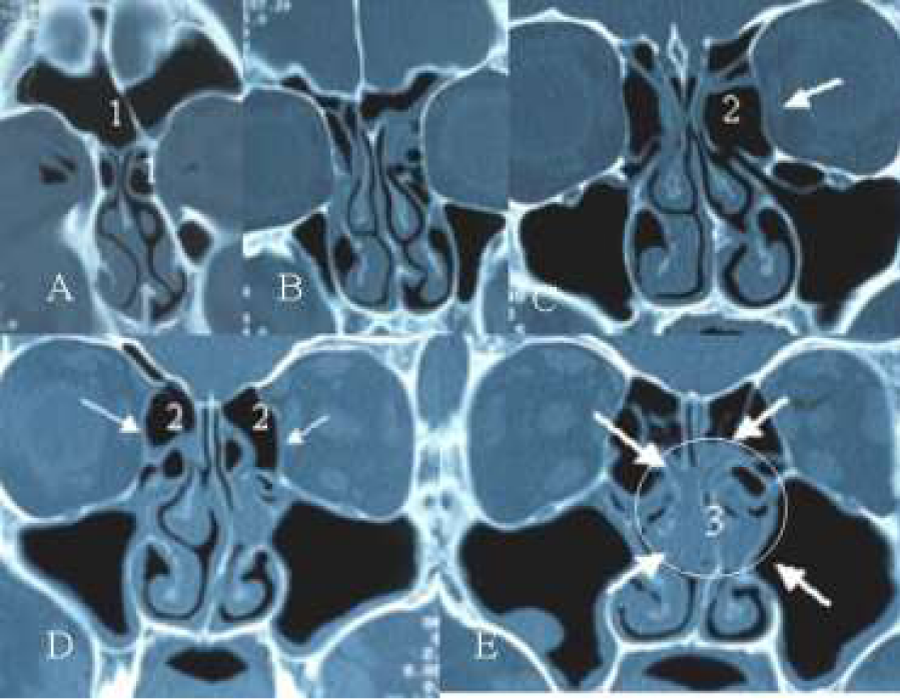
Figure 5:
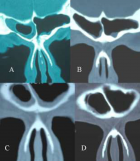
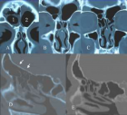
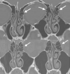
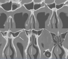
(A-E) Axial CT-scan showing presence of bilateral agger nasi cell (A), hyperinflated bulla ethmoidalis (C2) ballooning of the lamina orbitalis (C and D) and presence of an infra-orbital cell at the left side (C). Note also the diseased mucosa of both ethmoidal infundibulae and around both bullae leading to a beginning of a “black halo” aspect of the disease (E3) on the CT-scan.
Read Full Article HTML DOI: 10.29328/journal.hor.1001013 Cite this Article Read Full Article PDF
More Images
Similar Articles
-
CT signs of pressure induced expansion of paranasal sinus structuresPeter AR Clement*,Stijn Halewyck. CT signs of pressure induced expansion of paranasal sinus structures. . 2017 doi: 10.29328/journal.hor.1001013; 1: 077-087
Recently Viewed
-
A rare case of giant ovarian serous cystadenoma presenting as psuedo-meigs syndromeRichmond Ronald Gomes*,Sayeda Noureen,Habiba Akhter. A rare case of giant ovarian serous cystadenoma presenting as psuedo-meigs syndrome. Clin J Obstet Gynecol. 2021: doi: 10.29328/journal.cjog.1001078; 4: 010-014
-
Three visionaries for HRTJoseph Loze Onwude*. Three visionaries for HRT. Clin J Obstet Gynecol. 2021: doi: 10.29328/journal.cjog.1001077; 4: 007-009
-
The Utility of Acupuncture in Sports Medicine: A Review of the Recent LiteratureMichael Malone*. The Utility of Acupuncture in Sports Medicine: A Review of the Recent Literature. J Sports Med Ther. 2017: doi: 10.29328/journal.jsmt.1001004; 2: 020-027
-
Sinonasal Myxoma Extending into the Orbit in a 4-Year Old: A Case PresentationJulian A Purrinos*, Ramzi Younis. Sinonasal Myxoma Extending into the Orbit in a 4-Year Old: A Case Presentation. Arch Case Rep. 2024: doi: 10.29328/journal.acr.1001099; 8: 075-077
-
Sex after Neurosurgery–Limitations, Recommendations, and the Impact on Patient’s Well-beingMor Levi Rivka*, Csaba L Dégi. Sex after Neurosurgery–Limitations, Recommendations, and the Impact on Patient’s Well-being. J Neurosci Neurol Disord. 2024: doi: 10.29328/journal.jnnd.1001099; 8: 064-068
Most Viewed
-
Evaluation of Biostimulants Based on Recovered Protein Hydrolysates from Animal By-products as Plant Growth EnhancersH Pérez-Aguilar*, M Lacruz-Asaro, F Arán-Ais. Evaluation of Biostimulants Based on Recovered Protein Hydrolysates from Animal By-products as Plant Growth Enhancers. J Plant Sci Phytopathol. 2023 doi: 10.29328/journal.jpsp.1001104; 7: 042-047
-
Sinonasal Myxoma Extending into the Orbit in a 4-Year Old: A Case PresentationJulian A Purrinos*, Ramzi Younis. Sinonasal Myxoma Extending into the Orbit in a 4-Year Old: A Case Presentation. Arch Case Rep. 2024 doi: 10.29328/journal.acr.1001099; 8: 075-077
-
Feasibility study of magnetic sensing for detecting single-neuron action potentialsDenis Tonini,Kai Wu,Renata Saha,Jian-Ping Wang*. Feasibility study of magnetic sensing for detecting single-neuron action potentials. Ann Biomed Sci Eng. 2022 doi: 10.29328/journal.abse.1001018; 6: 019-029
-
Pediatric Dysgerminoma: Unveiling a Rare Ovarian TumorFaten Limaiem*, Khalil Saffar, Ahmed Halouani. Pediatric Dysgerminoma: Unveiling a Rare Ovarian Tumor. Arch Case Rep. 2024 doi: 10.29328/journal.acr.1001087; 8: 010-013
-
Physical activity can change the physiological and psychological circumstances during COVID-19 pandemic: A narrative reviewKhashayar Maroufi*. Physical activity can change the physiological and psychological circumstances during COVID-19 pandemic: A narrative review. J Sports Med Ther. 2021 doi: 10.29328/journal.jsmt.1001051; 6: 001-007

HSPI: We're glad you're here. Please click "create a new Query" if you are a new visitor to our website and need further information from us.
If you are already a member of our network and need to keep track of any developments regarding a question you have already submitted, click "take me to my Query."


