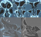
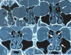
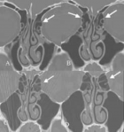
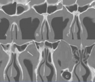
Figure 1
CT signs of pressure induced expansion of paranasal sinus structures
Peter AR Clement* and Stijn Halewyck
Published: 26 September, 2017 | Volume 1 - Issue 1 | Pages: 077-087
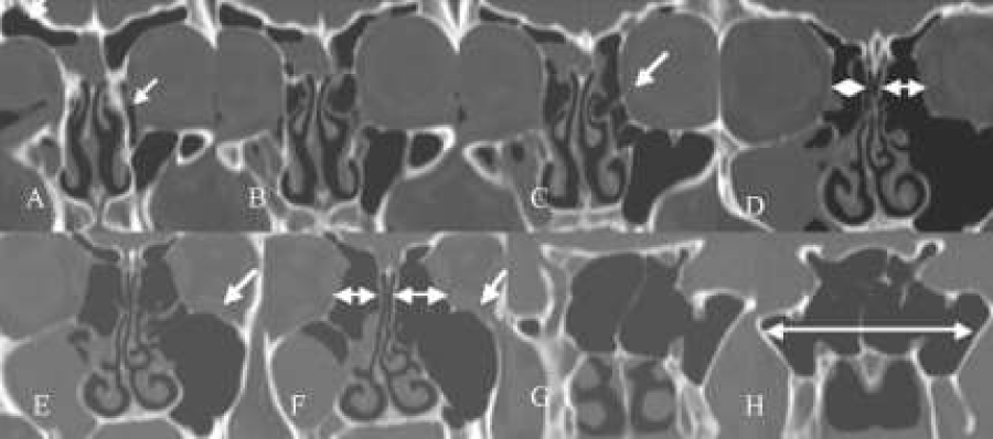
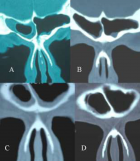
Figure 1:
(A-H) Coronal scans. Rounding of the supra-orbital recess (A,B), [ethmoidal emphysema of both sides but mainly on the left side, (ballooning lamina orbitalis and empty ethmoid - nearly no bony lamellae in the ethmioid itself)] very pneumatised bulla on the left (B,C,D) and on the right (E). Note different size between right and larger left ethmoid (E,F), waving of the orbital floor (E,F), and very extended sphenoidal sinus (G,H).
Read Full Article HTML DOI: 10.29328/journal.hor.1001013 Cite this Article Read Full Article PDF
More Images
Similar Articles
-
CT signs of pressure induced expansion of paranasal sinus structuresPeter AR Clement*,Stijn Halewyck. CT signs of pressure induced expansion of paranasal sinus structures. . 2017 doi: 10.29328/journal.hor.1001013; 1: 077-087
Recently Viewed
-
Intelligent Design of Ecological Furniture in Risk Areas based on Artificial SimulationTorres del Salto Rommy Adelfa*, Bryan Alfonso Colorado Pástor*. Intelligent Design of Ecological Furniture in Risk Areas based on Artificial Simulation. Arch Surg Clin Res. 2024: doi: 10.29328/journal.ascr.1001083; 8: 062-068
-
PTM-Fetuin-A: A Novel Biomarker for Early Detection of Diabetic Kidney DiseaseBenkova M,Petrov P,Aleksandar Petrov*,Staykova S,Zlatanova E,Chervenkov T,Nikolova S. PTM-Fetuin-A: A Novel Biomarker for Early Detection of Diabetic Kidney Disease. J Clini Nephrol. 2025: doi: 10.29328/journal.jcn.1001146; 9: 013-019
-
A rare case of giant ovarian serous cystadenoma presenting as psuedo-meigs syndromeRichmond Ronald Gomes*,Sayeda Noureen,Habiba Akhter. A rare case of giant ovarian serous cystadenoma presenting as psuedo-meigs syndrome. Clin J Obstet Gynecol. 2021: doi: 10.29328/journal.cjog.1001078; 4: 010-014
-
Three visionaries for HRTJoseph Loze Onwude*. Three visionaries for HRT. Clin J Obstet Gynecol. 2021: doi: 10.29328/journal.cjog.1001077; 4: 007-009
-
The Utility of Acupuncture in Sports Medicine: A Review of the Recent LiteratureMichael Malone*. The Utility of Acupuncture in Sports Medicine: A Review of the Recent Literature. J Sports Med Ther. 2017: doi: 10.29328/journal.jsmt.1001004; 2: 020-027
Most Viewed
-
Evaluation of Biostimulants Based on Recovered Protein Hydrolysates from Animal By-products as Plant Growth EnhancersH Pérez-Aguilar*, M Lacruz-Asaro, F Arán-Ais. Evaluation of Biostimulants Based on Recovered Protein Hydrolysates from Animal By-products as Plant Growth Enhancers. J Plant Sci Phytopathol. 2023 doi: 10.29328/journal.jpsp.1001104; 7: 042-047
-
Sinonasal Myxoma Extending into the Orbit in a 4-Year Old: A Case PresentationJulian A Purrinos*, Ramzi Younis. Sinonasal Myxoma Extending into the Orbit in a 4-Year Old: A Case Presentation. Arch Case Rep. 2024 doi: 10.29328/journal.acr.1001099; 8: 075-077
-
Feasibility study of magnetic sensing for detecting single-neuron action potentialsDenis Tonini,Kai Wu,Renata Saha,Jian-Ping Wang*. Feasibility study of magnetic sensing for detecting single-neuron action potentials. Ann Biomed Sci Eng. 2022 doi: 10.29328/journal.abse.1001018; 6: 019-029
-
Pediatric Dysgerminoma: Unveiling a Rare Ovarian TumorFaten Limaiem*, Khalil Saffar, Ahmed Halouani. Pediatric Dysgerminoma: Unveiling a Rare Ovarian Tumor. Arch Case Rep. 2024 doi: 10.29328/journal.acr.1001087; 8: 010-013
-
Physical activity can change the physiological and psychological circumstances during COVID-19 pandemic: A narrative reviewKhashayar Maroufi*. Physical activity can change the physiological and psychological circumstances during COVID-19 pandemic: A narrative review. J Sports Med Ther. 2021 doi: 10.29328/journal.jsmt.1001051; 6: 001-007

HSPI: We're glad you're here. Please click "create a new Query" if you are a new visitor to our website and need further information from us.
If you are already a member of our network and need to keep track of any developments regarding a question you have already submitted, click "take me to my Query."


