Figure 2
A comparative study of single or dual treatment of theranostic 188Re-Liposome on microRNA expressive profiles of orthotopic human head and neck tumor model
Shan-Ying Wang, Liang-Ting Lin, Bing-Ze Lin, Chih-Hsien Chang, Chun-Yuan Chang, Min-Ying Lin and Yi-Jang Lee*
Published: 25 February, 2021 | Volume 5 - Issue 1 | Pages: 001-012
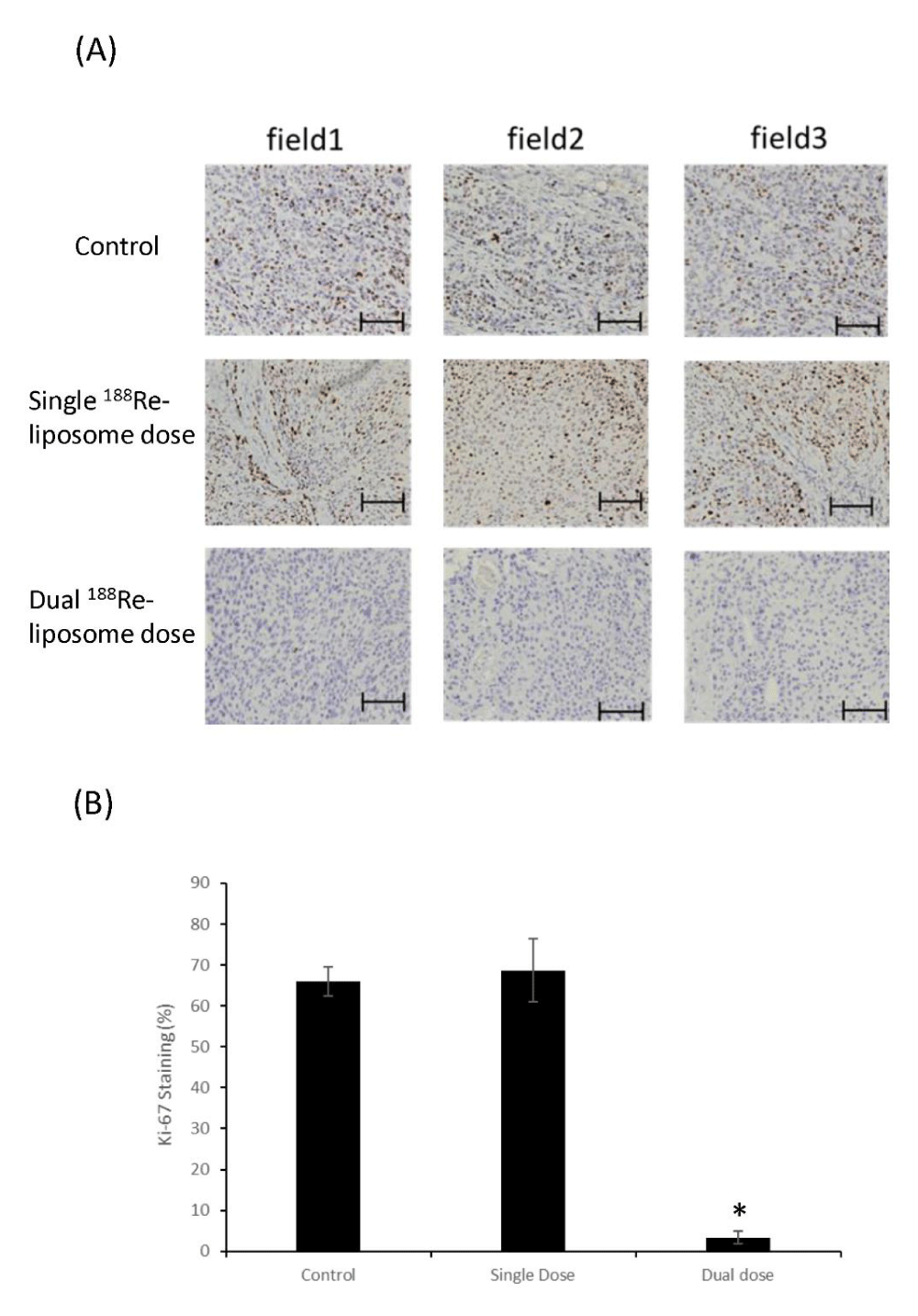
Figure 2:
Comparison of Ki-67 proliferative marks in HNSCC tumor sections. (A) IHC staining of Ki-67 in tumor sections with or without the treatment of 188Re-liposome. Three random fields were selected for imaging acquisition and quantification. Scale bar: 100 m. (B) Quantification of IHC staining of Ki-67 markers in tumor treated with different regimes of 188Re-liposome. *: p < 0.05.
Read Full Article HTML DOI: 10.29328/journal.hor.1001024 Cite this Article Read Full Article PDF
More Images
Similar Articles
-
A comparative study of single or dual treatment of theranostic 188Re-Liposome on microRNA expressive profiles of orthotopic human head and neck tumor modelShan-Ying Wang,Liang-Ting Lin,Bing-Ze Lin,Chih-Hsien Chang,Chun-Yuan Chang,Min-Ying Lin,Yi-Jang Lee*. A comparative study of single or dual treatment of theranostic 188Re-Liposome on microRNA expressive profiles of orthotopic human head and neck tumor model. . 2021 doi: 10.29328/journal.hor.1001024; 5: 001-012
Recently Viewed
-
A study of coagulation profile in patients with cancer in a tertiary care hospitalGaurav Khichariya,Manjula K*,Subhashish Das,Kalyani R. A study of coagulation profile in patients with cancer in a tertiary care hospital. J Hematol Clin Res. 2021: doi: 10.29328/journal.jhcr.1001015; 5: 001-003
-
Viral meningitis in pregnancy: A case reportRuth Roseingrave*,Savita Lalchandani . Viral meningitis in pregnancy: A case report. Clin J Obstet Gynecol. 2020: doi: 10.29328/journal.cjog.1001063; 3: 121-122
-
Vaginal and endometrial metastasis of primary cutaneous malignant melanomaMaria Boia Martins*,Francisca Morgado,Nuno Oliveira,Filomena Ramos. Vaginal and endometrial metastasis of primary cutaneous malignant melanoma. Clin J Obstet Gynecol. 2020: doi: 10.29328/journal.cjog.1001062; 3: 120-120
-
Universal testing for severe acute respiratory syndrome coronavirus 2 upon admission to three labor and delivery units in Santa Clara County, CASophia Yang,Rishi Bhatnagar,James Byrne,Andrea Jelks*. Universal testing for severe acute respiratory syndrome coronavirus 2 upon admission to three labor and delivery units in Santa Clara County, CA. Clin J Obstet Gynecol. 2020: doi: 10.29328/journal.cjog.1001060; 3: 109-113
-
Pregnancy complicated with deficiency of antithrombin: Review of current literatureMiroslava Gojnic,Zoran Vilendecic,Stefan Dugalic,Igor Pantic,Jovana Todorovic,Milan Perovic,Mirjana Kovac,Irena Djunic,Predrag Miljic,Jelena Dotlic*. Pregnancy complicated with deficiency of antithrombin: Review of current literature. Clin J Obstet Gynecol. 2020: doi: 10.29328/journal.cjog.1001059; 3: 103-108
Most Viewed
-
Evaluation of Biostimulants Based on Recovered Protein Hydrolysates from Animal By-products as Plant Growth EnhancersH Pérez-Aguilar*, M Lacruz-Asaro, F Arán-Ais. Evaluation of Biostimulants Based on Recovered Protein Hydrolysates from Animal By-products as Plant Growth Enhancers. J Plant Sci Phytopathol. 2023 doi: 10.29328/journal.jpsp.1001104; 7: 042-047
-
Sinonasal Myxoma Extending into the Orbit in a 4-Year Old: A Case PresentationJulian A Purrinos*, Ramzi Younis. Sinonasal Myxoma Extending into the Orbit in a 4-Year Old: A Case Presentation. Arch Case Rep. 2024 doi: 10.29328/journal.acr.1001099; 8: 075-077
-
Feasibility study of magnetic sensing for detecting single-neuron action potentialsDenis Tonini,Kai Wu,Renata Saha,Jian-Ping Wang*. Feasibility study of magnetic sensing for detecting single-neuron action potentials. Ann Biomed Sci Eng. 2022 doi: 10.29328/journal.abse.1001018; 6: 019-029
-
Pediatric Dysgerminoma: Unveiling a Rare Ovarian TumorFaten Limaiem*, Khalil Saffar, Ahmed Halouani. Pediatric Dysgerminoma: Unveiling a Rare Ovarian Tumor. Arch Case Rep. 2024 doi: 10.29328/journal.acr.1001087; 8: 010-013
-
Physical activity can change the physiological and psychological circumstances during COVID-19 pandemic: A narrative reviewKhashayar Maroufi*. Physical activity can change the physiological and psychological circumstances during COVID-19 pandemic: A narrative review. J Sports Med Ther. 2021 doi: 10.29328/journal.jsmt.1001051; 6: 001-007

HSPI: We're glad you're here. Please click "create a new Query" if you are a new visitor to our website and need further information from us.
If you are already a member of our network and need to keep track of any developments regarding a question you have already submitted, click "take me to my Query."


